What is cell cycle?
 The cell cycle is a life cycle or the life cycle of a cell. In eukaryotic cells (with a defined nucleus), the cell cycle is divided into interphase and M phase (mitosis or meiosis and cytokinesis).
For most of the time of a cell cycle, the cell is at the interface, being the preparatory, dormant part. The interface is divided into 3 stages: what is the cell cycle in biology
- The G 1 phase (presynthetic period): where the cell can remain for hours, days, or throughout its life,
- The S phase (synthesis period): where chromosomes replicate, and
- The G 2 phase: when the duplicate content is prepared for cell division.
On the other side, the “M” phases are divided into cytokinesis, where the cytoplasm is divided, and mitosis which is summarized in the following phases or processes: what is the cell cycle in biology
- Prophase : chromosomes condense, the mitotic spindle is created that captures the chromosomes, the nucleolus disappears and the nuclear envelope decomposes.
- Metaphase : the metaphysical plate is generated.
- Anaphase : sister chromatids are separated.
- Telefase : mitotic spindle disappears and the nucleolus appears. what is ell cycle in bigy
A cycle is characterized by not being linear. In this sense, each of the daughter cells has the ability to start the process again.
The cell cycle is important as the life cycle since they allow the reproductions and regeneration of the cells that make up all the organs, tissues, and elements of living organisms.
What’s on this page? what is cell cycle in biology
 Stage of cell division
- DNA replication
- Mitosis
- Stage of mitosis
- Mitosis: late
- Mitosis: the end stage
- Cell cycle summary
Stages of cell cycle what is cell cycle in biology
- G in the G1 and G2 phases represents Gap. It is indicated that there are no significant changes in the nucleus during these two phases ; in fact, these cells are very active, they are growing and are preparing for division.
- S in the S phase represents synthesis. At this stage, the DNA is replicated.
- M in the M phase represents mitosis , at which stage one cell divides into two daughter cells. what is cell cycle in biology
To help you understand cell division more intuitively, here’s an animation of this cell division process, and the same animation is shown at the end of the page.
Many anti-tumor drugs work by blocking one or more links in the cell cycle. In order to better understand the defects that occur in tumor cells and the mechanism of action of anti-tumor drugs used to block cell division, we will study the cell cycle in more detail. what is cell cycle in biology
DNA replication what is cell cycle in biology
DNA replication occurs during the synthesis phase of the cell cycle (ie, S phase). With the participation of many enzymes (enzymes) of each chromosome ( chromosome ) are replicated with a high degree of accuracy. In this process, the double helix structure of DNA is released, and each single strand replicates another complementary strand as a template. The end result is that the genetic material forms two identical “copyes”.
The animation below shows this process.
The copied chromosome contains two identical DNA strands. The two chains remain attached until they are separated by mitosis. Since this form of chromosome is the easiest to identify and most intuitive, most people are familiar with it. The figure below shows the process of chromosome replication.

It should be noted that the X-shaped structure in the above figure is actually two “copy copies” copied from one chromosome.l cycle in biology
During the copying process, an error may occur, resulting in a change in the nucleotide sequence of the chromosome . If this change occurs in genes, it will affect the function of the cell. Human cells have evolved several mechanisms to correct such errors, but these mechanisms are not perfect. Errors in DNA replication can lead to the appearance of mutated gene cells, and the accumulation of these variability can lead to malignant tumors. The occurrence of several malignant tumors is associated with the failure of the repair process involved in normal DNA replication. The process of generating variation will be discussed in the section “Causes of Variation”. what is ell cycle in biology
All cells that are dividing must undergo a DNA replication process. Since cancer cells are often rapidly dividing, so this phase of the cell cycle became many chemotherapy ( Chemotherapy ) of targets. This will be discussed in the chapter “Cancer Treatment”. Such chemotherapy drugs include doxorubicin, cyclophosphamide, carboplatin, cisplatin and etoposide. well cycle in biology
Detailed introduction of chromosomes and genes
Most of the DNA in the cell is in the form of chromosomes in the nucleus. The human body has 46 chromosomes, each pair consisting of a pair of 23 pairs. Parents pass 23 chromosomes to the next generation through gametes (sperms and eggs). That is, the parent passes one of each pair of chromosomes (such as one of the first pair, one of the second pair, one of the third pair, …) to the next generation. This means that each person gets 23 chromosomes from their parents, so there are 23 pairs in total. Each chromosome consists of a single DNA strand. In the DNA strand, millions of nucleotides bind to several different proteins. The gene is distributed on the chromosome with a large number of unclear DNA. well cycle in biology
Every gene always appears in the same position on the same chromosome. For example, if the gene that determines the color of the eye is located on chromosome 1 of a person, then all the genes of other people examined are also on chromosome 1. on. Since the chromosomes we have are all paired, this means that each gene has two “copy copies.” The figure below depicts this relationship. The chromosome marked with “arrow” represents the chromosome from the father, and the chromosome marked with “ten” represents the chromosome from the mother. It is important to know that the ” versions ” of genes in each pair of chromosomes do not need to be the same. Continue to discuss the above example. The gene from the father’s eye color determines that the eyes appear blue, while the same gene from the mother makes the eyes brown. Then, the color of the child’s eyes is determined by the result of the interaction of the two copies of the gene. what ill cycle in biology
In the following screen, the color bands represent genes. For some genes, the “version” of the genes from both parents is the same; however, for some genes, there is a slight difference. Stripes of different colors indicate different gene “versions” (ie alleles). The pair of chromosomes shown in the figure below represent two “versions” of the same chromosome from both parents (eg, pair 1, pair 2, pair 3, …). what is cell cycle in biology
Mitosis what is the cell cycle in biology
 The most studied cell cycle is the M phase, the mitosis phase. During the mitosis phase, a single cell divides into two daughter cells. In normal cells, mitosis produces two daughter cells that contain the same genetic material as the mother cells. As we will observe, tumor cells do not always follow this pattern. Mitosis can be further divided into several stages depending on changes occurring within the cell (especially changes in the nucleus of the nucleus).
The first stage is the prophase of mitosis . At this stage, the nuclear envelope dissolves and the chromosomes (chromosomes) concentrate to prepare for cell division. Just like the entanglement on the bobbin, such concentration makes the chromosomes denser, making it easier for chromosomes to be distributed into the two emerging daughter cells. Also, in early mitosis, protein ( Protein ) fibers (i.e., spindle fiber spindle fibers) is formed and connected to the bipolar cells. This fiber bundle acts as a scaffold for the cells in the division, and the cell needs this scaffold to push and pull the cellular components to form two daughter cells. what is cell cycle in biology
The bundle of protein fibers that connect the two poles of the cell is called a microtubule. These proteins are aggregated and depolymerized during cell division. They have become targets for a variety of chemotherapy. Taxol® is a chemical extracted from yew trees that binds to microtubules and prevents microtubules from depolymerizing. In this way, the cells are unable to complete mitosis and die. Another type of chemotherapeutic drug is represented by vinblastine, which acts just the opposite, blocking the aggregation of the spindle. But the final effect is the same as that of paclitaxel, which inhibits cell division. To learn more, see the chapter on the treatment of malignant tumors.
Detailed introduction of human chromosomes
The figure below shows the entire chromosomes of human cells (chromosomes). The method of describing all chromosomes in this way is called karyotype (ie, karyotype ). The karyotype is often used to detect abnormalities in the chromosomes of the fetus in pregnant women. These chromosomes are stained by binding to fluorescent dyes. It should be noted that all chromosomes are paired. There are significant differences in the size of the chromosomes. Biologists number them in descending order. Chromosome 1 is the largest, and chromosomes 21 and 22 are the smallest. The karyotype shown below is from a male, including an X chromosome and a smaller Y chromosome. Even the smallest chromosomes, the DNA inside contains millions of base pairs. what is cell cycle in biology

In many malignant cells, the number of chromosomes is affected, manifested as too many or too few. Cells containing too many or too few chromosomes are called aneuploid cells. For more information, see Gene Variations and Tumors.
Image courtesy of Santa Clara, CA.
Stage of mitosis what is cell cycle in biology
The figure below shows cells in the prophase of mitosis. A concentrated, X-shaped chromosome can be seen in the figure.

Each chromosome is actually composed of two identical DNA strands. The number of these DNAs doubled as early as the S phase of cell division. We will continue to discuss the S phase of cell division soon.
 In the next stage of the mitosis phase, chromosomes are arranged in the equatorial plate in the middle of the cell and are distributed in equal amounts to the two daughter cells.
Mitosis: late
The two equal parts of the chromosome are dragged to the ends of the cell to form two daughter cells; the two daughter cells are identical to the mother cells. In the following two stages of mitosis (ie, late and late stages), the cells will complete chromosome segregation and cell division. what is cell cycle in biology

The figure below shows the chromosomes in the dividing epithelial cells of the rat kangaroo. This is a chromosome image that is dragged to the poles of the cell at the end of mitosis.
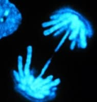
The above image is licensed by the copyright holder Molecular Probes®.
Mitosis: the end stage

At the end of mitosis, the nuclear membrane reforms, marking the completion of cell division. The figure below shows a periodic cell division process. The “interval” is the interval between two cell divisions during which the cells  exert their physiological effects in the body. Most of the time, the cells are in the “interval”.
The following is an animated illustration of the cell division process:
Cell cycle summary
Introduction what is cell cycle in biology
- Over time, many of the cells that make up the body age and die, and new cells are needed to replace them.
- The process by which cells divide to produce two identical copies is called mitosis.
- Cells produced by mitosis are called daughter cells.
- The cell division process is a four-stage ordered process, collectively referred to as the cell cycle.
- The abnormal shape of many cancer cells is due to defects in genes that control cell division.
cell cycle
- The cell cycle consists of four phases: G1, S, G2, and M.
- The G1 and G2 phases are intervals (Gap), and cells in the interval grow and are ready to divide.
- The chromosome (DNA) in the S phase is replicated.
- The mitosis is a stage of cell division in which cells divide into two daughter cells.
- Most cells do not actively divide, and these cells are at rest (G phase).
Mitosis (M phase)
- In normal cells, mitosis forms two cells with identical genetic information.
- There are four sub-stages of mitosis:
- Pre-phase – chromosome agglutination, chromosomal nuclear membrane decomposition, spindle formation
- Mid-term – the copied chromosomes are arranged in the cell midline (equatorial plate)
- Late – chromosome separation, the cells grow longer and form two segments (two poles)
- Terminal – nuclear membrane re-formed at both ends, as the new cell membrane is formed into two separate cells
DNA synthesis (S phase)
- Humans have 46 chromosomes and parents provide 23 pairs.
- Each chromosome consists of a strand of DNA consisting of millions of nucleotides.
- Homologous chromosomes have the same genes, but the “versions” of genes are different.
- In many cancer cells, the number of chromosomes changes, resulting in too many or too few chromosomes in the cell. These cells are called aneuploidy.
- Errors may occur during DNA replication, leading to genetic variations that may be carcinogenic.
- Cells have mechanisms to modify DNA replication errors.
- Many chemotherapeutic drugs are treated for the S phase of the cell cycle.