We explain what a microscope is, when and who invented this instrument. The parts that compose it and types of microscope.
-
What is a microscope?
The microscope is an instrument that allows you to observe objects that are too small to be seen by the sight of the human being . The term microscopes is the conjunction of two concepts, on the one hand ” micro ” that is equivalent to ” small ” and ” scopio ” which means “to observe “, in short it refers to small observation , or to a lesser extent.
The microscope is an optical instrument that increases the ability to observe at approach levels such that particle analysis is even possible . The image that is obtained is really an investigation on the composition of the objects. The study and analysis of small objects is called ” microscopy .”
-
When and who invented the microscope?

This instrument was invented by Zacharias Janssen in the year 1590 . The discovery of this instrument was very important, mainly for its contributions in medical research. In 1665, the research conducted by William Harvey on blood circulation appeared, when analyzing blood capillaries. In 1667, Marcello Malpighi, an Italian biologist, was the first researcher to study living tissues thanks to observation through a microscopes.
The Dutchman Anton van Leeuwenhoek used microscopes to describe for the first time various organisms , protozoa , bacteria , sperm and red blood cells . It can be considered as the founder of science that studies the behavior of bacteria , gave rise to bacteriology. The innovative thing about his technique is that he carried out the studies with his own microscopes, spent much of his time in shaping magnifiers, giving the crystals the millimeter thickness he needed.
From there on, more technical progress has been made by increasing the level of magnification of microscopes, and this in turn enabling medical science to carry out more and more thorough research on the behavior of microorganisms and cell studies . The progress thanks to the implementation and development of the microscopes was enormous in the 18th century.
Then came the electron microscopes, developed in Germany in 1931 by two researchers Max Knoll and Ernst Ruska. This made it possible to achieve an increase of 100,000X, a huge leap for the technique.
-
Parts of a microscope
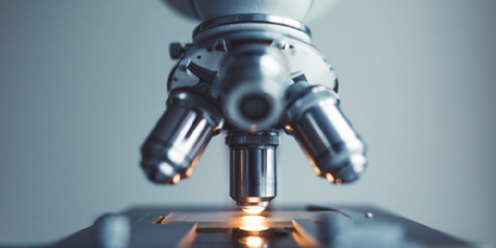
The different parts that make up a microscopes commonly are:
- Eye lens It is where you place the eye of the observer. This lens can increase the image between 10 to 15 times its size.
- Canyon. It is basically an elongated metal tube whose interior is black, serves as a support for the eyepiece and objective lenses.
- Objective lenses. It is a group of 2 or 3 lenses located in the revolver.
- Stir. It is a system that contains the objective lenses inside, it can have a rotation system that allows the exchange of these lenses.
- The macrometric screw. It is a knob that rotates it by zooming in or out of the object being observed.
- The micrometric screw. It is what allows to refine and focus the image correctly. Making it clearer.
- The stage It is a clamp platform, where the object or the preparation you want to observe is placed.
- The diaphragm It serves to regulate the amount of light that passes through the object under observation.
- The condenser It serves to concentrate the light beam in the preparation or object.
- Artificial light source. directs light to the stage.
-
Types of microscopes
There are various types of microscopes that were used throughout history , and there are also currently designed microscopes with a special purpose, some of these are:
- Scanning electron microscopes
- Optical microscopes
- Simple microscopes
- Compound microscopes
- Ultraviolet light microscopes
- Fluorescence microscopes
- Petrographic Microscopes
- Dark field microscopes
- Phase contrast microscope
- Polarized light microscope
- Confocal microscope
- Electronic microscope
- Transmission electron microscope
- Ion Microscope in Field
- Scanning probe microscope
- Tunnel effect microscope
- Atomic force microscope
- Virtual microscope


